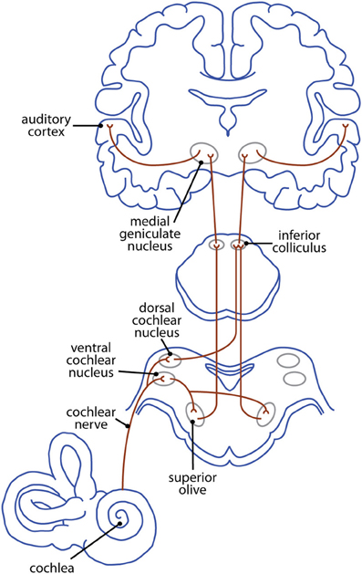
The physiology of the cochlea is considered elsewhere and this section follows the auditory pathway from the organ of Corti to the Auditory cortex. Afferent neurones arising from the basilar membrane send their information to the cochlear nuclei in the medulla. The pathways to the primary auditory cortex in the middle third of the superior temporal gyrus, are complex and unlike the other sensory pathways in that they are biilateral, i.e. information from each cochlea is relayed through both sides of the brainstem to both suditory cortices, left and right. There are some important relay stations including the superior olivary nuclei of the medulla, the inferior colliculi in the midbrain, and the medial geniculate nucleus of the thalamus. As for the visual pathway, the colliculi of the midbrain are concerned with eye movements. The superior colliculi are conerned with directing the gaze towards object in the visual field. The inferior colliculi are concerned with directing the gaze - turning the eyes -towards objects that are identified in auditory space. One of the factors that is involved in determining the origin of sounds is the time difference between the arrival of sounds ant the two ears, normally meausred in a millisecond or two. The auditory pathway travels up both sides of the brainstem, and that allows the system to identify precise timings of activity in the two ears at an early stage of the pathway. The superior olivary nucleus seems to be concerned with detecting this interaural time difference; it is believed that the difference in the volume of sound reaching both ears may also be detected in this part of the pathway.. The medial geniculate body is concerned with relaying the frequency and intensity of sounds to the auditory cortex; information concerning the timing and intensity of information arising from both ears is also relayed to the cortex. There seem to be maps of sound frequencies within the inferior colliculi and the medial geniculate body (tonotopic maps, which correspond to the somatotopic map in the somatosensory cortex and the maps of the visual fields in the visual cortices). The auditory cortex in each temporal lobe recieves information for both ears and analyses the frequency volume and interaural timing of sound. Neurons at one end of the auditory cortex respond best to low frequencies; neurons at the other respond best to high frequencies. These different parts of the auditory area are concerned therefore with the pitch and volume of a sound, while other sub-areas are concerned with musical sounds, composed of a fundamental frequency and its harmonics. |
 * *
Primary Auditory Cortex The primary auditory cortex is essential for the perception of sounds, but central deafness is rare in humans because both cortices receive similar information, and it is rare for both to be injured by lesions.
Eye Movements in relation to the direction of sound The inferior colliculi are concerned with directing the gaze towards objects that are identified in auditory space. Lower parts of the auditory pathway can still function however if the perception of sound is lost, so eye movements in response to sound can still occur. |
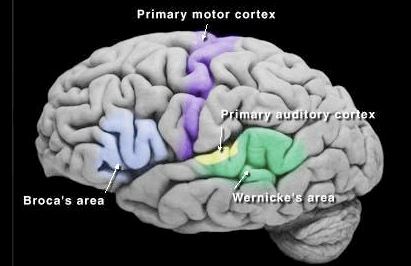
Wernicke's area is one of the two parts of the cerebral cortex associated the the understanding of the spoken word. It is located in the posterior section of the superior temporal gyrus behind the primary auditory area. Destruction to Wernicke's area results in an inability to understand commands, written or spoken, known as a sensory dysphasia, or sensory aphasia. The area receives its input from the primary auditory area, and sends its output to Broca's area, through the arcuate fasciculus. Broca's area is concerned with speech, and lesions of this area or the arcuate fasciculus are associated with a motor dysphasia, in which the patient can understand commands, but cannot form spoken words, or has slurred speech and words that are imperfectly formed. There is some uncertainty as to the exact anatomical site of Wernicke's and Broca's areas, but the speech recognition area is though to be in the Sylvian fissure near the junction of the temporal and parietal lobes, and the arcuate fasciculus may project to the pre-motor cortex. However the classical identities of these functions are still in common use as descriptions of the functions associated with each area. |
In most people Broca's and Wernicke' areas are found only in the left cerebral hemisphere. |
The development of language skills depends on areas of the parietal lobe that develop late-on in human development. The Arcuate Fasciculus, a large C-shaped bundle of axons that connect Wernicke's area and Broca's area, and areas adjacent. There is no clear definition of these two classical areas involved in speech recognition and vocal responses, and it seems likely that there are a variety of subdivisions of cortical functions within these areas. |
 * *
Plasticity in Hearing and Speech In most adults the areas of the brain concerned with speech recognition and vocalisation are in the left hemisphere. However in young children who sustain damage to the left hemisphere, the right side can take over these functions. There are examples of children who have had surgical removal of epileptic foci from the left hemisphere, who go on to develop normal speech functions using the right hemisphere. This suggests that the neuroplasticity in the right hemisphere takes over functions that are more commonly found in the left hemisphere. |

